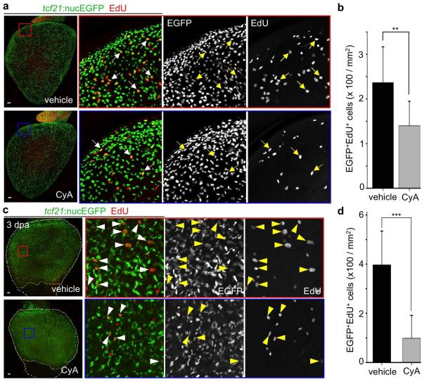
Extended Data Figure 8. Epicardial proliferation is regulated by Hh signaling.
a, Freshly dissected tcf21:nucEGFP hearts were randomly separated into two groups and cultured for 47 hours with vehicle (n = 11) or 5 μM CyA (n = 8). Then, 25 μM EdU was added to the medium for one hour prior to collection at 48 hours. Cyclopamine (CyA) treatment decreases epicardial cell proliferation ex vivo. Arrows, representative EGFP+ (Green) EdU+ (Red) nuclei. b, Quantification of EGFP+EdU+ nuclei per mm2 on the ventricular surface, from hearts in (a). **P < 0.01; Student's two-tailed t-test. c, tcf21:nucEGFP adult fish were subjected to partial ventricular resection surgery, and randomly separated into two groups for treatment with vehicle (n = 8) or 10 μM cyclopamine (CyA) (n = 10) from 2 to 3 dpa. Then, 10 mM EdU was injected intraperitoneally 1 hour prior to collection. CyA treatment decreases epicardial cell proliferation in vivo. Arrowheads, representative EGFP+ (Green) EdU+ (Red) nuclei. d, Quantification of EGFP+EdU+ nuclei per mm2 on the ventricular surface, from hearts in (c). ***P < 0.001; Mann-Whitney Rank Sum Test. Yellow dashed lines in (c), resection plane. White dashed lines in (c), ventricle. Boxed areas in (a, c), magnified views. Scale bars, 50 μm. Error bars, s.d.

