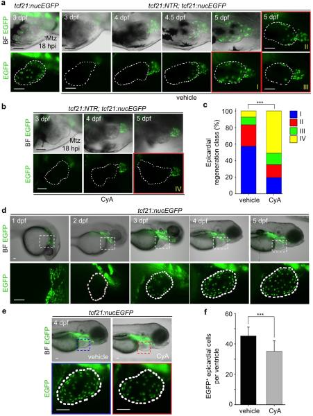
Extended Data Figure 9. Larval epicardial development and regeneration.
a, tcf21:nucEGFP or tcf21:NTR; tcf21:nucEGFP larval clutchmates were treated with 10 mM Mtz from 6 hpf to 54 hpf, and then imaged at different times from 3 to 5 dpf. tcf21:nucEGFP larvae show normal ventricular epicardial coverage at 3 dpf, while tcf21:NTR; tcf21:nucEGFP coverage is sparse. tcf21:NTR; tcf21:nucEGFP larvae with confirmed full ablation were imaged from 3 to 5 dpf, covering first the ventricular base and then the apex. Three different extents of regeneration at 5 dpf are shown: classes I, greater than two-thirds coverage; II, one-third to two-thirds coverage; and III, some cells but less than one-third coverage. b, A subset of tcf21:NTR; tcf21:nucEGFP larvae with confirmed full ablations were randomly separated and treated with vehicle or CyA, which limited regeneration in most cases (class IV, no ventricular epicardial cells). c, Quantification of extents of regeneration from experiments in (a) and (b). ***P < 0.001; Chi-square test; n = 54 embryos for vehicle, 51 for CyA. d, Epicardial morphogenesis visualized in tcf21:nucEGFP larvae. No epicardial cells are evident at or before 2 days post-fertilization (dpf). By 3 dpf, ventricles contained 17.6 +/- 6 epicardial cells on average (n = 23), whereas 4 dpf larvae contained 45.2 +/- 5.8 cells (n = 21). e, tcf21:nucEGFP larval clutchmates were randomly separated into two groups for treatment with vehicle or 5 μM CyA from 2 to 4 dpf. f, Quantification of ventricular EGFP+ epicardial cells from groups in (e). ***P < 0.001; Student's two-tailed t-test; n = 21 for each group. White dashed lines in (a, b, d, e), ventricle. Boxed areas in (d, e), magnified views. Scale bars, 50 μm. Error bars, s.d.

