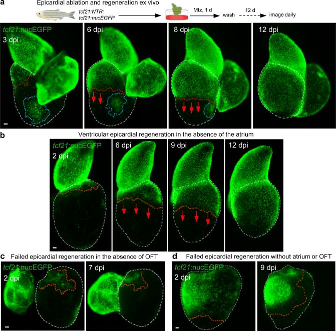
Figure 2. Cardiac outflow tract is required for regeneration of adjacent ventricular epicardium.
a, (Top) Schematic for epicardial ablation and regeneration in hearts cultured ex vivo. (Bottom) Regeneration occurs in a base-to-apex direction (arrows). Isolated patches (circled by blue dashed lines) do not participate in regeneration until contacted by the leading edge. b, Ventricular epicardium regenerates in the absence of the atrium (n = 19; behavior seen in all samples). Arrows, direction of regeneration. c, d, Ventricular epicardium fails to regenerate in the absence of the BA. Ventricular epicardium showed defective regeneration in these experiments with (c) (n = 6; all samples) or without atrium (d) (n = 14; all samples), even when, in rare cases, many basal epicardial cells were spared (d). Red dashed lines, epicardium or epicardial leading edge. White dashed lines, ventricle. Scale bars, 50 μm.

