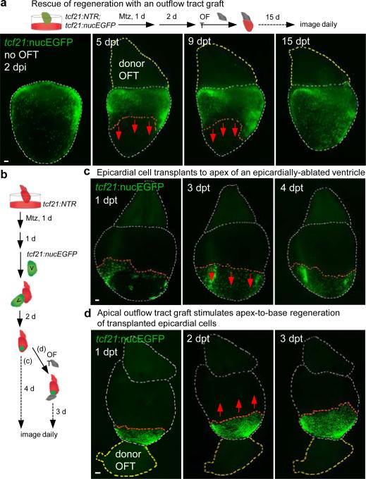
Figure 3. Outflow tract tissue is sufficient to initiate and redirect epicardial regeneration.
a, (Top) A non-transgenic BA (donor OFT) was transplanted to a transgenic ventricular base after epicardial ablation ex vivo. (Bottom) Base-to-apex epicardial regeneration (arrows) was observed from host tissue in 13 of 21 ventricles. dpt, days post-transplantation. b, (Top) Experimental design in (c, d). c, tcf21:nucEGFP+ epicardial cells transplanted to an epicardially ablated ventricular apex were static or regenerated (arrows) toward the apex (n = 12, all samples), but not toward the base. d, tcf21:nucEGFP+ epicardial cells were transplanted to the apex of an epicardially-ablated ventricle, followed by apical grafting of a donor BA. tcf21:nucEGFP+ cells regenerated in a reversed apex-to-base direction (arrows) in 9 of 14 ventricles. Twelve of 18 host ventricles with the host BA removed before donor BA grafting also showed apex-to-base regeneration. Red dashed lines in (a, c, d), epicardial leading edge. White dashed lines in (a, c, d), ventricle and host BA. Yellow dashed lines in (a, d), donor BA. Scale bars, 50 μm.

