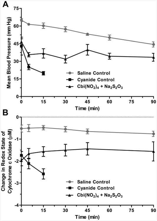
Figure 6.

Nitrocobinamide and thiosulfate as a cyanide antidote in rabbits. New Zealand white rabbits were anesthetized and received a continuous intravenous infusion of sodium cyanide. When their MAP was <70% of precyanide infusion values, generally after ∼30 min of cyanide infusion, the animals received an intramuscular injection of saline (cyanide control, black squares) or antidote [Cbi(NO2)4 plus Na2S2O3, black triangles]. The time of injection is defined as zero time. In separate experiments, rabbits were anesthetized and treated similarly, except they received a continuous intravenous infusion of saline (saline control, gray circles) instead of sodium cyanide and they did not receive an intramuscular injection. (A) MAP is plotted versus time. (B) Cytochrome c oxidase redox state is plotted versus time. The data are the mean ± SD of three independent experiments.
