Figure 1.
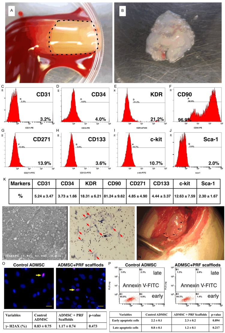
Preparation of platelet-rich fibrin (PRF) scaffold and engineered adipose-derived mesenchymal stem cell (ADMSC) grafts and flow cytometric analysis of stem cells at day 14 cell culturing. A. The PRF scaffold, white jelly-like component, (black dot-line square) was already prepared in the Eppendorff tube. B. The example of one piece of engineered ADMSC grafts had been prepare and already for patching on the infarct area. C to J. The illustrat ion of flow cytometric expressions of endothelial progenitor cells (EPCs) and ADMSCs at day-14 cell culture in DMEM culture medium. K. The mean distributions (n = 6) of EPC (CD31, CD34, KDR, CD133) and ADMSC (CD90, CD271, Sca-1, c-Kit) surface markers. CD90 (i.e., ADMSC) was the highest population among the culturing stem cells. L. The scanning electronic microscopy clearly identified the morphological feature of the homogeneous and network-like PRF scaffold with the attachment of numerous ADMSCs (black arrows) in the surface layer of the scaffold. M. Under microscopic finding (100 ×), numerous DAPI stained ADMSCs (red arrows) were identified at the bottom layer of the transwell membrane after 24 h migration. N. Illustrating the PRF scaffold (black dotted line) which was covered on the surface of infarct area and was sutured (black arrows) for fixation. O. The immunofluorescent microscopic findings (400 ×) showed no difference in γ-H2AX+ cells (yellow arrows) between with and without PRF scaffold treatment. Scale bars in right lower corners represent 20 μm. P. The flow cytomtric analysis displayed no difference in early or late apoptosis of ADMSC between with and without PRF scaffold treatment.

