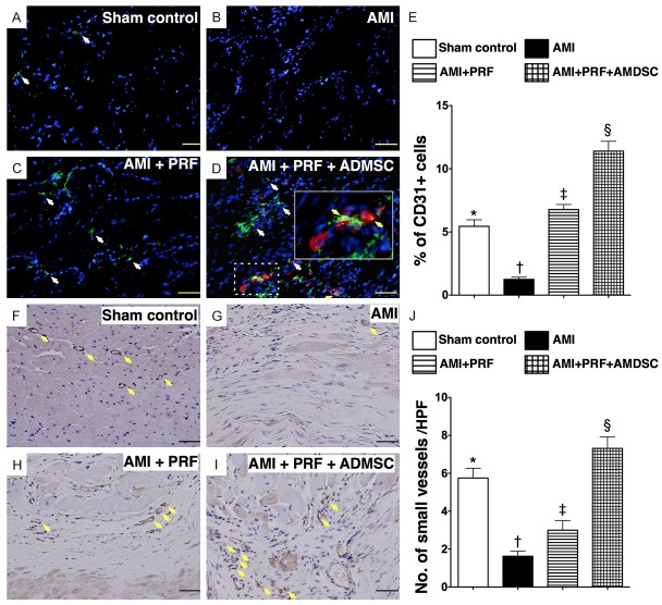
Figure 4.
Immunofluorescent (IF) and immunohistochemical (IHC) staining for endothelial cells and small vessels on day 42 after AMI induction (n = 8). (A to D) IF microscopic (200 ×) findings of CD31+ cells (white arrows) in infarct area (IA) of LV myocardium. Red color (D) indicated the Dil-dye stained ADMSCs migrated into the LV myocardium from epicardial engineered ADMSC grafts. Merged image (D) from double staining (Dil-dye + CD31) demonstrating cellular components with mixed red-green color (yellow arrows) under high magnifications (600 ×) (solid-line square from dot-line squared which being magnified), indicating some of engineered ADMSC grafts differentiated into endothelial cells (CD31+). Scale bars in right lower corners represent 50 μm. (E) *vs. other groups with different symbols (*, †, ‡, §), p < 0.0001. (F to I) IHC staining (α-smooth muscle actin staining) in infarct area of LV myocardium for identification of small vessels (< 15 μm) (yellow arrows) microscopically (200 ×) in four groups. Scale bars in right lower corners represent 50 μm. (J) *vs. other groups with different symbols (*, †, ‡, §), p < 0.0001. HPF = high-power field. Statistical analysis for (E) and (J) using one-way ANOVA, followed by Bonferroni multiple comparison post hoc test. Symbols (*, †, ‡, §) indicate significance (at 0.05 level).

