
Figure 5.
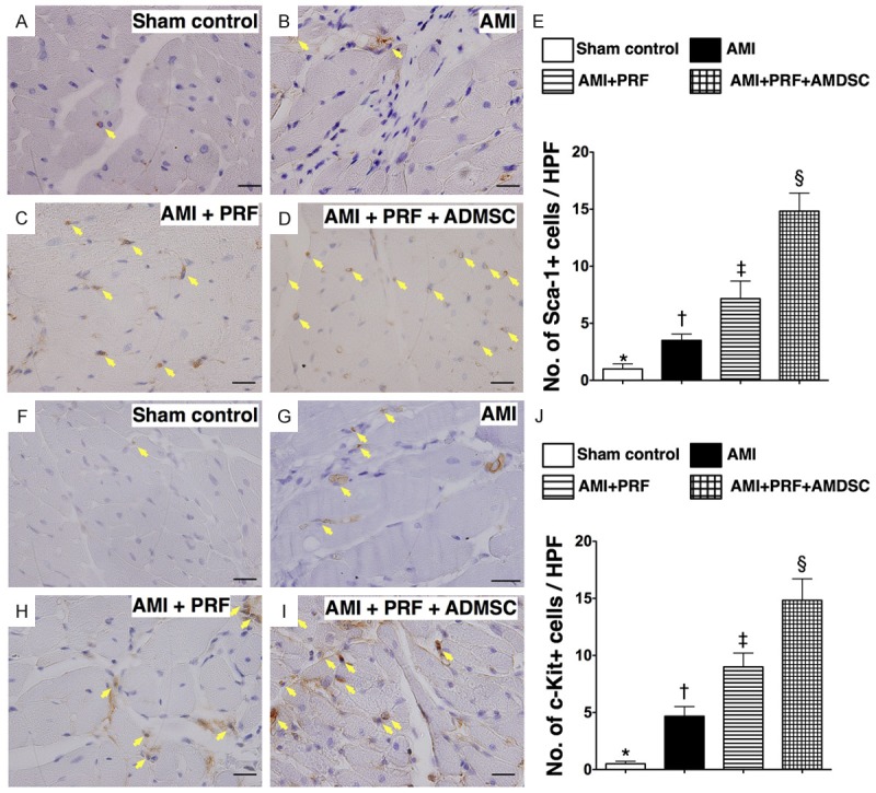
Immunohistochemical (IHC) staining for identification of mesenchymal stem cellsin infarct area of LV myocardium on day 42 after AMI induction (n = 8). (A to D) IHCmicroscopic(200 ×) findings of Sca-1+ cells (yellow arrows) in LV myocardium. (E) *vs.other groups withdifferent symbols (*, †, ‡, §), p < 0.0001. (F to I) IHC microscopic(200 ×) findings ofc-Kit+ cells (yellow arrows) in LV myocardium. (J) *vs. other groupswith different symbols(*, †, ‡, §), p < 0.0001. HPF = high-power field. Statisticalanalysis for (E) and (J) using one-way ANOVA,followed by Bonferroni multiple comparisonpost hoc test. Symbols (*, †, ‡, §) indicate significance (at 0.05 level).