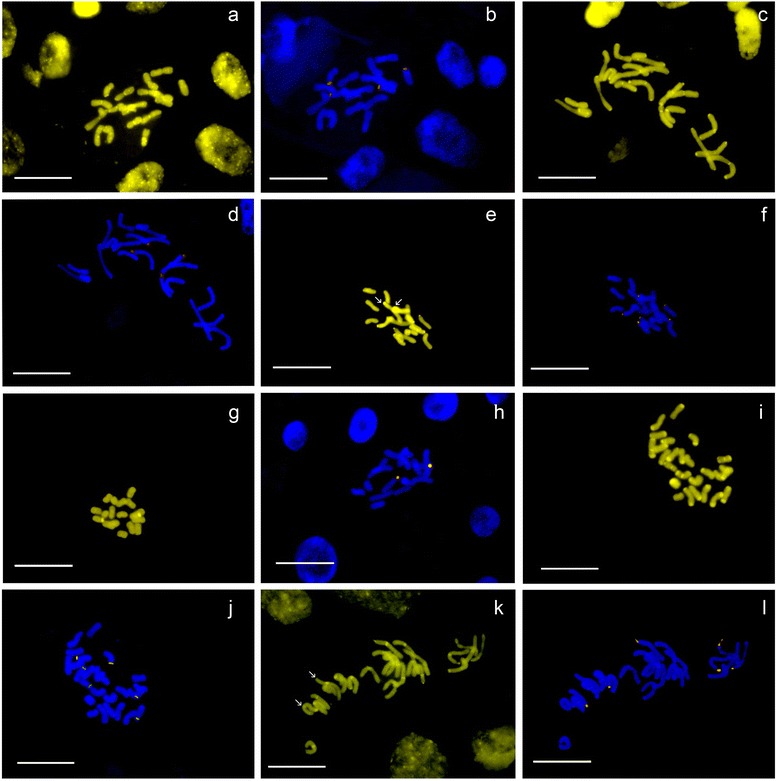
Fig. 3.

Chromomycin A3-positive (CMA+) FISH images of cytogenetically variable Tanacetum species, in which CMA+ bands are marked yellow, 26S-5S rDNA signals and marked orange. (a, b) T. archibaldii (2x) with 56 CMA signals (asterisks indicate interacalary CMA+ bands) and with 4 rDNA signals; (c, d) T. balsamita, 2x, with 40 CMA+ signals (many of them pericentromeric, indicated with asterisks) and with four rDNA signals – a slightly decondensed rDNA is indicated with an arrow; cultivated (e, f) and wild (g,h) T. parthenium (from Shahid Beheshti University, 1633 and Tochal, 1483, respectively), both 2x with 14 and six CMA+ and six and two rDNA signals observed, respectively; (i, j) T. kotschyi (Tabriz, Mishodagh, 1339), 3x, with 44 CMA+ signals and six rDNA signals and (k, l) T. joharchii, 3x, with 24 CMA and six rDNA signals; note faint or interstitial CMA+ bands indicated with asterisks and decondensed rDNAs indicated with arrows in both pictures. Scale bars = 10 μm
