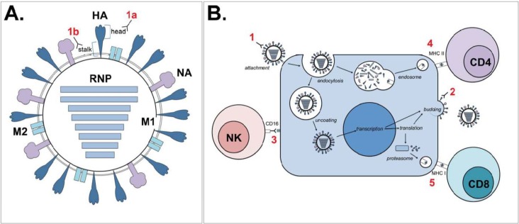
Figure 1.
Schematic representation of possible immunological correlates of protection. Numerals in red show various immunological correlates of protection as indicated below. (A) Cartoon of an influenza virion, showing the hemagglutinin (HA) surface glycoprotein (stem and head), the neuraminidase (NA) surface glycoprotein, the matrix 2 (M2) ion channel, the matrix 1 (M1) structural protein and the ribonucleoproteins (RNPs: the combination of genomic RNA, viral polymerases PA, PB1 and PB2 and nucleoproteins (NP)). Antibodies directed against HA can either target the globular head (1A) or stem region (1B). (B) Interference of production of progeny virus by infected cells by various immunological correlates of protection, including (1) antibodies against HA head, interfering with binding or HA stem, potentially interfering with post-entry functions of HA, like endosomal membrane fusion; (2) antibodies against NA, limiting the production of progeny virus; (3) antibodies against M2e, HA or NA, followed by ADCC through CD16 signaling in NK cells (or phagocytosis, not shown); (4) virus-specific CD4+ T lymphocytes; and (5) virus-specific CD8+ T lymphocytes that possess cytolytic activity.

