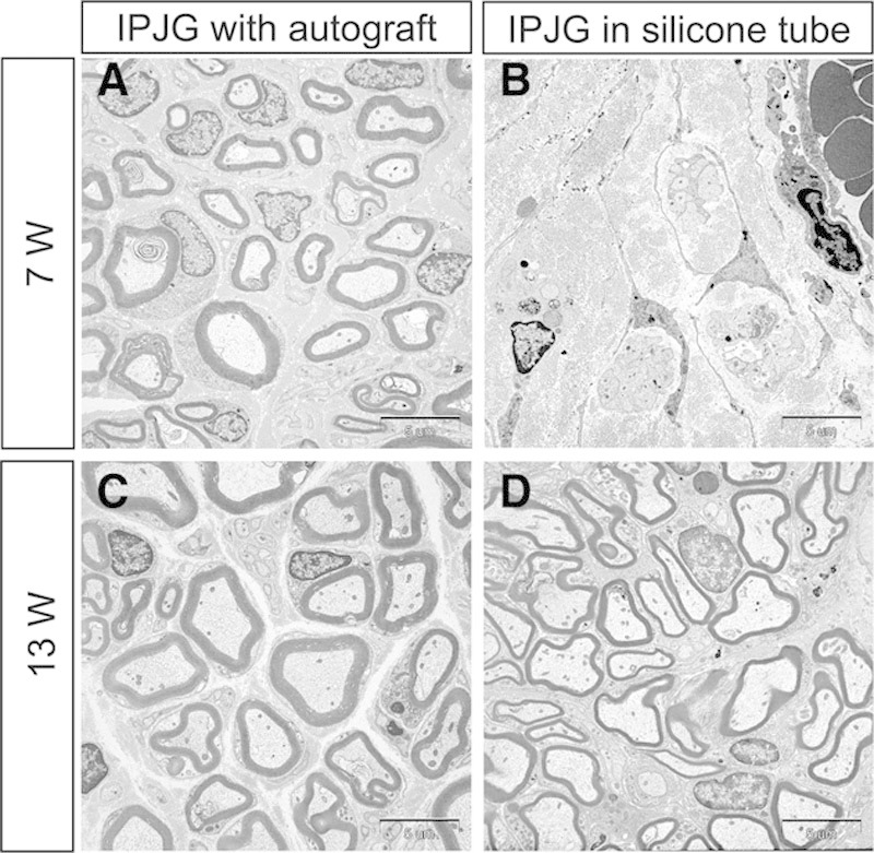
Fig. 4.

Transmission electron microscope observations of IPJGs at postoperative weeks 7 (A and B) and 13 (C and D). The left and right columns show IPJGs with autograft and in a silicone tube conduit, respectively. The upper and lower rows show the observations at postoperative weeks 7 and 13, respectively. B, Although axonal regeneration was observed in the regenerating nerve in the silicone tube group at week 7, no regeneration of the myelin sheath was observed. D, A comparatively thick and substantial annular myelin sheath was observed in the silicone tube group at week 13. A and C, Regeneration of mature myelin sheath was observed in the autograft group at postoperative week 7 and 13.
