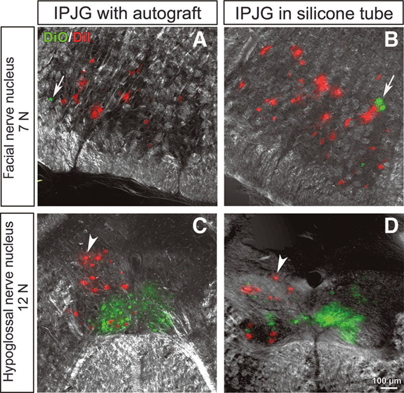
Fig. 7.

Retrograde tracer findings of IPJGs at postoperative week 13 in the autograft (A and C) and silicone tube (B and D) groups. DiI and DiO, fluorescent retrograde tracers, were injected into the whisker pads and tongue, respectively. The left and right columns show IPJGs with autograft and in silicone tube conduit, respectively. The upper and lower rows show the observations at facial nerve nucleus (7N) and hypoglossal nucleus (12N), respectively. A, DiO-positive (green) motor neurons (white arrow) were found to be mixed with DiI-positive (red) motor neurons in 7N of the autograft group. C, In the hypoglossal nerve nucleus, DiI-positive (red) motor neurons (white arrowhead), which were mixed with DiO-positive (green) motor neurons, had an irregular arrangement (white arrowhead). B, Similarly, a mixture of DiI-positive (red) motor neurons and DiO-positive (green) motor neurons (white arrow) in 7N were found in the silicone tube group. D, In the hypoglossal nerve nucleus, DiI-positive (red) motor neurons (white arrowhead) mixed with DiO-positive (green) motor neurons had an irregular arrangement. The results confirmed the double innervation of the facial muscles of expression and the tongue by 7N and 12N. Unusual labelings after IPJG treatments were indicated with white arrows and white arrowheads.
