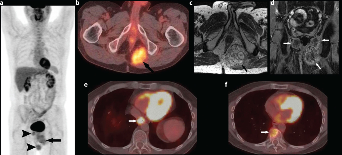
Figure 1. a–f.
47 year-old male presented with low abdominal pain: Coronal image from F-18 FDG PET/CT shows three FDG avid masses, the largest one in the left ischiorectal fossa (arrow). Two smaller masses (arrowheads) are also seen adjacent to it and on the right suggesting recurrent disease (a). Axial image from F-18 FDG PET/CT shows FDG avid mass in the left ischiorectal fossa (arrow) (SUV (Standardized uptake value) Max 4.8) (b). Axial FSE T2 sequence shows large septated ill-defined T2 hyperintense mass in the ischiorectal fossa (arrow) (c). Coronal post contrast sequence shows heterogeneous enhancement in bilateral ischiorectal fossa (arrows) (d). Axial image from F-18 FDG PET/CT shows intense FDG uptake associated with a mass in the distal esophagus (SUV max 25) (arrow), consistent with biopsy proven oesophageal primary malignancy (e). Axial image from F-18 FDG PET/CT shows FDG-avid (SUV 5.6) lytic lesion involving T10 vertebral body (arrow), highly suspicious for metastatic disease (f).

