
Figure 2. a–c.
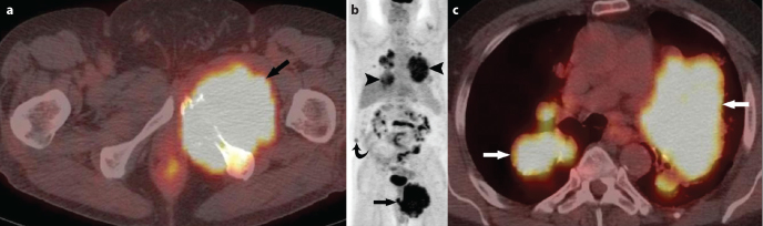
58 year-old man presented with pelvic pain: Axial image from F-18 FDG PET/CT shows a large FDG-avid mass in the left hemipelvis (SUV Standardized uptake value) 12.9) (arrow) with the destruction of the inferior pubic rami (a). Coronal image from F-18 FDG PET/CT shows large FDG-avid mass in the left hemipelvis with a SUV max of 12.9 (arrow), multiple FDG-avid pulmonary nodules/masses with a SUV max of 8.3 (arrowheads), mildly FDG avid soft tissue nodule in the right abdominal wall (SUV max 3.4) suspicious for metastatic implant (curved arrow) (b). Axial image from F-18 FDG PET/CT. Multiple FDG-avid pulmonary nodules/masses (arrows) are seen with a SUV max of 8.3 (c).