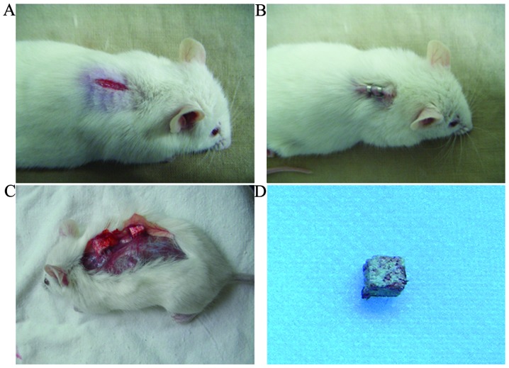
Figure 5.
Procedures of the animal study. Six-week-old female SCID mice were used for the subcutaneous transplantation study. (A) Midlongitudinal skin incisions of ~1 cm in length were made on the dorsal surface of each mouse, and subcutaneous pockets were generated by blunt dissection. (B) Each animal was implanted with four three-dimensional porous ceramic graft transplants containing human dental pulp cells incubated with 1.8 or 5.4 mM Ca2+, with one single transplant per pocket. The incisions were closed with surgical staples. (C and D) Implants were harvested at day 7 after transplantation, fixed for 1 day in 4% formaldehyde in phosphate-buffered saline, and the cell growth and mineralization were examined by scanning electron microscopy.

