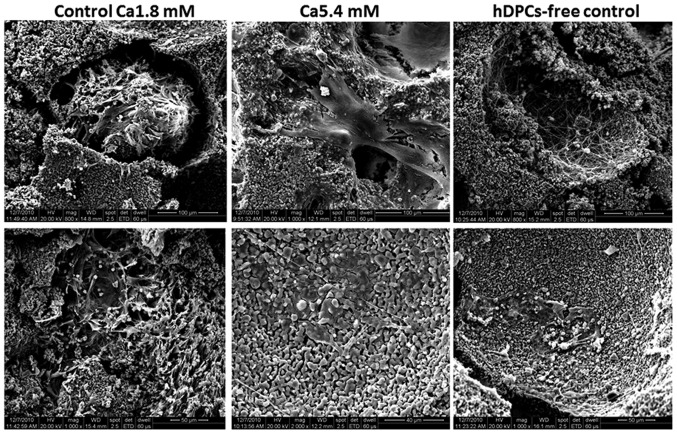
Figure 6.
Scanning electron microscopy images of hDPC-HA/TCP complexes harvested from SCID mice (scale bar: Top row, 100 µm; bottom row, 50 µm). In the control Ca1.8 mM HA/TCP scaffolds, a large number of hDPCs proliferated significantly and aggregated to form a dense multilayer with clumping. By contrast, hDPCs cultured in medium containing 5.4 mM Ca2+ displayed undefined cell borders and intercellular fusion, and numerous extracellular matrixes on the scaffolds’ surface were formed. hDPC; human dental pulp cell; HA/TCP, hydroxyapatite/tri-calcium phosphate.

