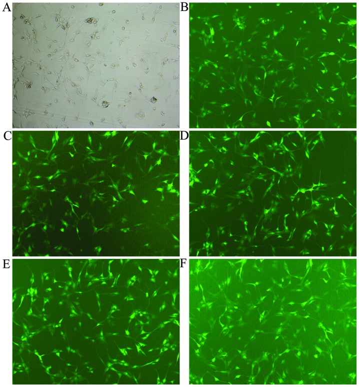
Figure 2.
The lentivirus (LV) transfection efficiency at different multiplicities of infection (MOIs), as shown under a fluorescence microscope (magnification, ×100). (A and B) Nucleus pulposus (NP) cells were observed under an inverted microscope and a fluorescence microscope at an MOI of 60. (C–F) LV transfection efficiency at 30, 40, 80 and 100 MOI under a fluorescence microscope, respectively.

