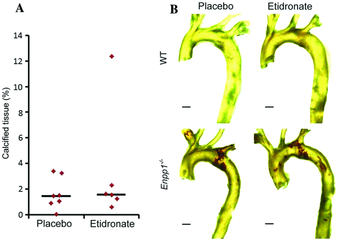
Figure 2.
(A) Quantification (% of calcification) of calcium deposition in the aortae of 22-week-old Enpp1−/− mice. A standardised region of calcium deposition (400 slices from the subclavian artery) was selected and revealed no significant differences between the placebo (vehicle; saline)- and etidronate-treated groups. (B) Three-dimensional volumetric reconstructions of aortae from 22-week-old wild-type (WT) placebo-treated, WT etidronate-treated, Enpp1−/− placebo-treated, and Enpp1−/− etidronate-treated mice. Calcification is indicated by brown colouring. Bar represents 0.5 mm.

