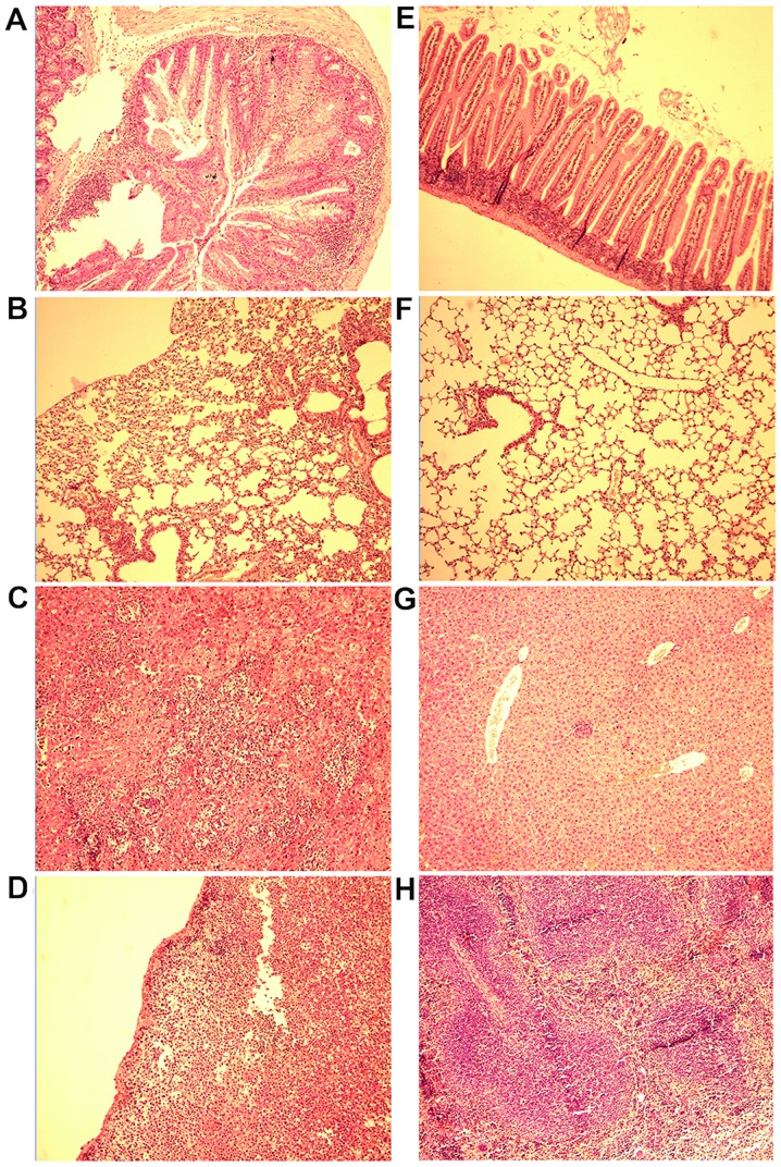
Figure 6.
Histological analysis of the mice injected with A20 cells alone and mesenchymal stem cell (MSC) plus A20 cells on day 28 following transplantation. Mice in the (A-D) A20 group and (E-H) MSC-A20 group were sacrificed, and the small intestine, lungs, liver, and spleen were removed, fixed, paraffin-embedded, and sectioned. Paraffin-embedded 5-µm tissue sections were stained with hematoxylin and eosin. (A) Small intestinal mucosa presented necrosis, defluxion and vacuolar degeneration. (B) Collapse of pulmonary alveoli. (C) Large tumor nodule infiltrating the liver, crushing normal liver tissues. (D) A20 cells infiltrated the spleen. (E-F) Normal small intestine and lung structures. (G-H) Normal liver and spleen structures. Objective lens: magnification, ×10.

