
Fig. 1. WRN-deficient MSCs exhibit phenotypes associated with premature cellular senescence.
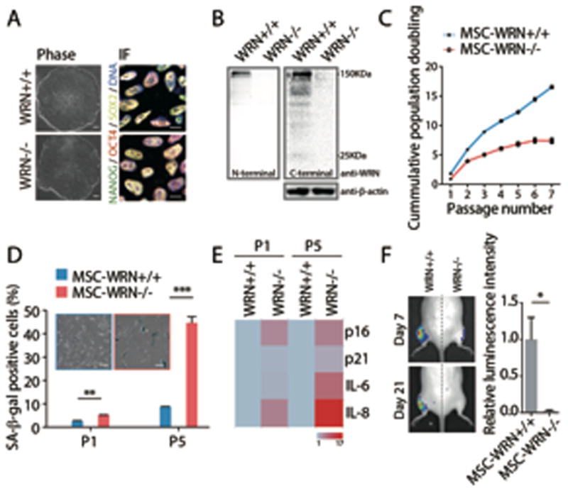
(A) Morphology and immunofluorescence analyses of pluripotency markers in ESCs. Scale bar, 100 μm and 10 μm, respectively. (B) Western blot analysis of WRN expression in ESCs using anti-WRN N-terminal (ab200) and C-terminal (SC-5629) antibodies. (C) Growth curve analyzing the accumulative population doubling of MSCs. (D) Senescence-associated (SA)-β-gal staining in passage 1 (P1) and P5 MSCs. Scale bar, 50μm. (E) Quantitative RT-PCR analysis of the indicated genes in P1 and P5 MSCs. Transcript levels were normalized to MSCs-WRN+/+ group. Genes with greater mean value are color coded towards red. (F) Photon flux from muscle of NOD-SCID mouse transplanted with MSCs-WRN+/+ (left) and MSCs-WRN−/− (right) expressing luciferase. All data are represented as mean + SEM. *P<0.05, **P<0.01, ***P<0.001 by t test; n=3.