
Fig. 3. WRN associates with centromeric heterochromatin, and forms a molecular complex with SUV39H1 and HP1α.
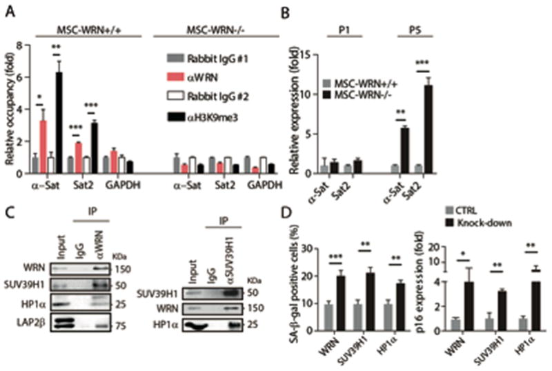
(A) Enrichment of WRN and H3K9me3 within the region of α-Sat or Sat2 as measured by ChIP-qPCR. (B) Quantitative RT-PCR analysis of centromeric repetitive element transcripts in MSCs at the indicated passages. (C) Left, co-immunoprecipitation of SUV39H1, HP1α, and LAP2β protein with endogenous WRN protein; Right, co-immunoprecipitation of WRN and HP1α with endogenous SUV39H1 in wild-type MSCs. (D) SA-β-gal staining (left) and p16 transcript (right) analyses in wild-type MSCs transduced with control lentiviral vector (CTRL) or lentiviral vector encoding for the indicated shRNA (Knock-down). All data are represented as mean + SEM. *P<0.05, **P<0.01, and ***P<0.001 by t test; n=3.