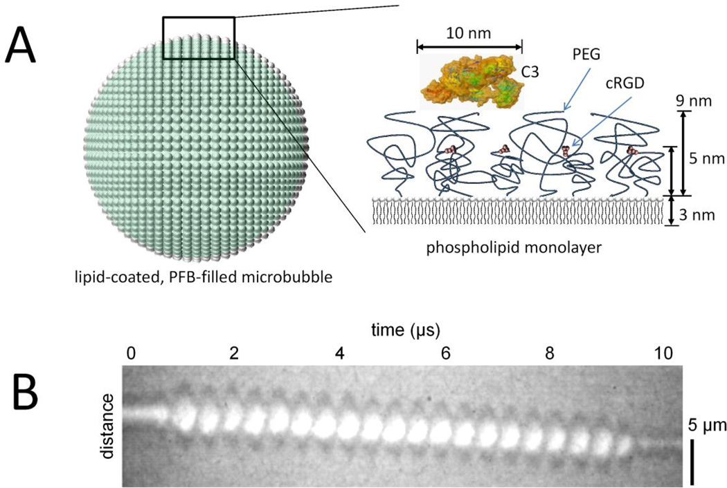
Figure 1.
Buried-ligand microbubbles and ultrasound radiation force. A, The ultrasound contrast agent is a microbubble of 4 to 5 µm diameter filled with perfluorobutane (PFB) gas and coated with a phospholipid monolayer shell. A side view of the shell shows the buried-ligand architecture, which comprises a shorter (2,000 Da) polyethylene glycol (PEG) tethered to cyclo Arg-Gly-Asp (cRGD) peptide surrounded by a longer (5,000 Da) PEG overbrush.5–9 Also shown is a surface rendering of complement C3 protein.27 B, A high- speed videomicroscopic streak image of the diameter of a microbubble over time shows acoustic radiation force acting on a microbubble to displace it axially away from the ultrasound transducer superimposed with surface dilations. These surface dilations expose the cRGD for binding to the target αVβ3 integrin.

