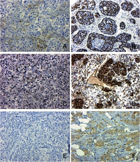
Fig. 4.

Negative immunoperoxidase staining for cyclin D1, FOXP1 and NRP1 in BRCA1 basal cancers (a, b and e). Positive staining in sporadic basal cancers for cyclin D1, FOXP1 and NRP1 (b, d and f). Note positive nuclear staining in stromal cells and negative staining in tumour cells for cyclin D1 (a) and FOXP1 (c). (x10, Haematoxylin counterstain)
