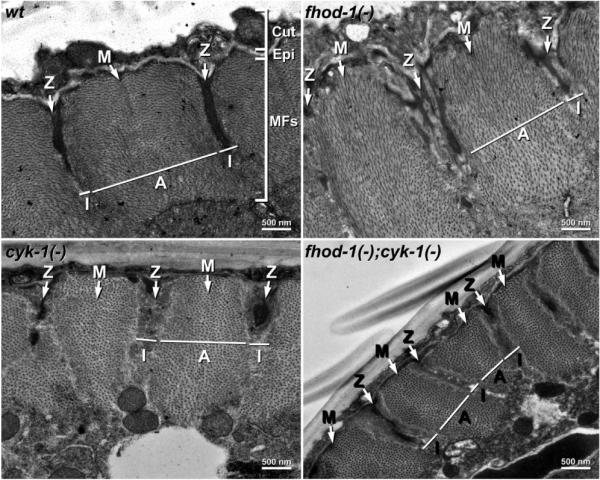
Figure 2.

TEM of wild-type and formin mutant sarcomeres. Cross section (x/y) views through the body walls of young adult wild-type and indicated formin mutant worms show the outer cuticle (Cut), the thin epidermis (Epi), and the thick layer of BWM myofilaments (MFs) organized into sarcomeres. The positions of example Z-lines, M-lines, A-bands, and I-bands are indicated.
