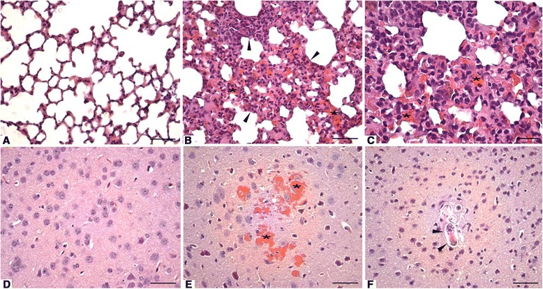
Fig. 5.

Lung and brain parenchyma of BALB/c mice infected with Toxocara canis. a Control group: normal lung parenchyma. H&E staining. Bar = 50 μm. b 14 days post-infection (p.i.): thickening of the septum due to inflammatory infiltrate (arrowheads) and hemorrhagic areas (*). H&E. Bar = 50 μm. c Higher magnification of the previous figure (14 days p.i.) showing inflammatory infiltrates consisting of eosinophils, lymphocytes, macrophages and the presence of hemorrhagic areas (*). H&E. Bar = 20 μm. d Control group: normal brain parenchyma. H&E. Bar = 20 μm. e 7 days p.i.: brain with hemorrhagic cavities (*). H&E. Bar = 20 μm. f 14 days p.i.: presence of larvae in the brain (arrowheads). H&E. Bar = 20 μm
