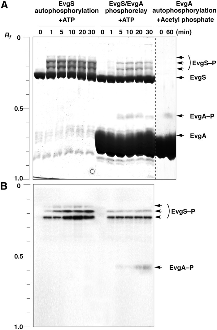
Fig 2. Profiling of EvgS autophosphorylation, the EvgS/EvgA phosphorelay, and EvgA autophosphorylation by using Phos-tag SDS-PAGE.
(A) The EvgS autophosphorylation and EvgS/EvgA phosphotransfer reactions were performed in the presence of 30 mM ATP. The EvgA autophosphorylation reaction was performed in the presence of 40 mM acetyl phosphate. Gels were stained with cCBB. The incubation times for the reactions are shown above each lane. Each lane contained 2 μg of EvgS or EvgA. (B) The EvgS autophosphorylation and EvgS/EvgA phosphotransfer reactions were performed in the presence of 370 kBq of [γ-32P]-ATP. The gel was subjected to autoradiography.

