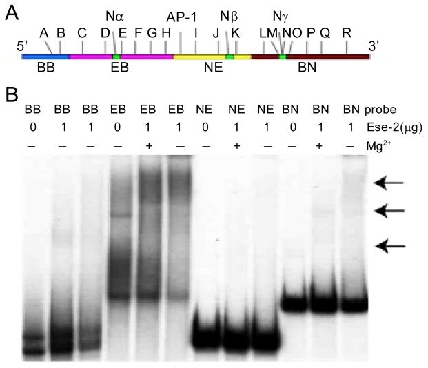
Fig. 2.
Full length Ese-2 cannot efficiently bind the first intron of K18. (A) Schematic diagram of the first intron of K18 showing the four 32P-labelled DNA fragments that span the intron. (B) Electrophoresis mobility shift assay using DNA fragments BB, EB, NE, and BN (1×10⁴ cpm each). Each DNA fragment was incubated with 1 μg full length Ese-2 protein that was expressed and purified from E. coli. Arrows indicate regions of possible shift with EB and BN DNA fragments.

