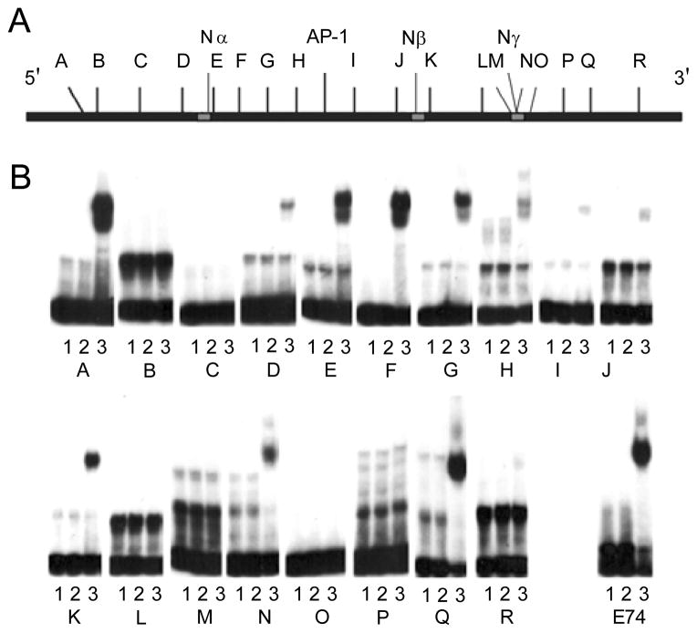
Fig. 4.
Ese-2-ets selectively binds EBSs within the first intron of K18. (A) Schematic diagram of the first intron of K18. Negative regulatory elements (Nα, Nβ, Nγ) are indicated in grey. EBS locations corresponding to oligonucleotides A-R used in gel shift experiments are indicated. (B) Electrophoresis mobility shift assays. Each double-stranded oligonucleotide (K18 oligonucleotides A-R or E74) was incubated with or without (see lane descriptions) 100 ng Ese-2-ets E. coli expressed and purified protein. Lane 1, oligonucleotide (1×10⁴ cpm) only; Lane 2, 100 ng wild type Ese-2; Lane 3, 100 ng Ese-2-ets.

