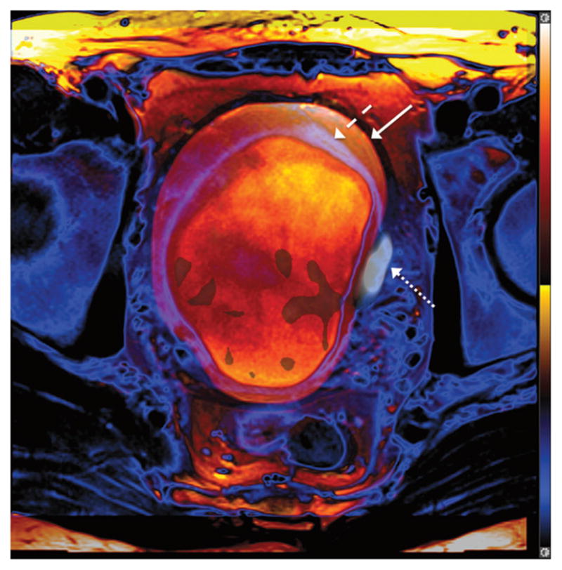
FIGURE 3.

A 52-year-old man with muscle-invasive high-grade bladder cancer. Fusion of 2 axial TSE T2WIs obtained at separate points during the PET/MRI examination, with the bladder wall shaded in purple (dashed arrow) and blue (solid arrow) for the early and later acquisitions, respectively. Bladder has expanded between the 2 acquisitions, as indicated by the outer positioning of the bladder wall in the later image. Left lateral bladder wall tumor exhibits increased 18F-FDG activity (dotted arrow) on PET obtained simultaneously as later T2WI. Tumor shows improved coregistration with simultaneous, rather than sequential, T2WI.
