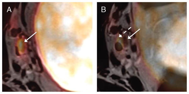
FIGURE 4.

A 52-year-old man with muscle-invasive high-grade bladder cancer (same patient as in Figure 2). A, Fused image from axial TSE T2WI and simultaneously acquired 18F-FDG PET acquisition demonstrates small right pelvic side wall lymph node with increased FDG activity (solid arrow). Registration of the lymph node on the 2 images is excellent. B, Fused image from sequentially obtained axial T2WI and PET demonstrates misregistration between lymph node on MRI (dashed arrow) and PET (solid arrow). Also note improved coregistration of bladder wall on simultaneous image.
