
Figure 5. Microfluidic chip migration assay of U87 glioblastoma cells.
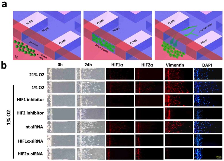
(a) Schematic diagram of the microfluidic chip migration assay. Cells are placed in a chamber containing either 21% or 1% oxygen and allowed to migrate along a three-dimensional collagen-based surface. (b) U87 glioma cell migration following exposure to 21% (top row) or 1% oxygen saturation for 24 hours. Immunofluorescent stains against HIF1α, HIF2 or vimentin are shown in red and DAPI is blue. Cells in the bottom five rows were treated with a HIF1α inhibitor, HIF2α inhibitor, non-targeting siRNA (nt-siRNA), HIF1α-siRNA or HIF2α-siRNA, as indicated. Hypoxia increased the distance and number of vimentin-positive cells that migrated onto the collagen surface. Total migration was significantly reduced after treatment with a HIF inhibitor and attenuated following HIF knockdown by siRNA. Treatment with a HIF2 inhibitor or siRNA knockdown of HIF1α or HIF2α significantly reduced the number of migrating vimentin-positive cells. Scale bar = 100 μm.