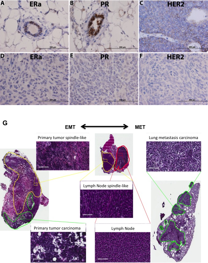
Figure 2. JygMC(A) mammary primary tumor molecular phenotype, histology and EMT-MET plasticity.

Analysis of normal adjacent tissue by immunohistochemistry of ERα A. PR B. and HER2/neu staining of a wild-type MMTV-Neu/Her2 tumor C. Analysis of primary mammary tumor tissue of ERα D. PR E. and HER2/neu F. Positive antibody signals are shown in brown, and the hematoxylin counterstain is shown in blue. Scale bars: 200μm. G. Histological tumor classification. Primary tumor possesses two types of cells: undifferentiated adenocarcinoma type (green line) and areas composed of atypical spindle-shaped cells suggesting EMT (yellow line). Lung metastasis morphology typical of the epithelial primary adenocarcinoma suggesting MET is present in the lung parenchyma. Lymph node tissue (red line) showing areas of atypical spindle-shaped cells (yellow line). Shown are hematoxylin and eosin staining. Scale bars: 100 μm.
