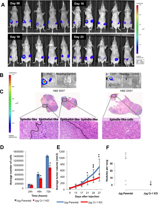
Figure 7. Mouse Cripto-1 promoter and Cripto-1 knockout in JygMC(A) cell line.

Representation of bioluminescent imaging on animals injected bilaterally into the fourth mammary gland with JygMC(A) cells containing the mouse Cripto-1 promoter. Animals were imaged at day 8, 16, 19 and 23-post cell injection. B. In situ detection of Cripto-1 promoter activity in primary tumor tissue sections. C. H&E of the primary tumor tissue sections depicted in B. showing carcinoma areas (green) and EMT-like areas (yellow) and high magnification (20X). Scale bars: 3mm and 200μm. D. Proliferation assay. JygMC(A) cells were seeded in 12-well dishes in triplicate at 5x10⁴cells/well and cultured for 24, 48 and 72 hrs. Cells were then harvested and counted. Data are representative of two independent experiments in triplicate ±SD, *P < 0.0002, as compared to control cells. E. Average of primary tumor volumes are represented in the graph on JygCr-1KO and JygMC(A) parental cells (n = 7 animals/group ±SD, *P < 0.01 and **P < 0.0001, as compared to control animals). F. Number of pulmonary nodules per animal in JygCr-1KO and JygMC(A) parental animals (*P < 0.05, one-sided values; Wilcoxon rank-sum test).
