
Figure 5. Establishment of a NSCLC bioluminescent mouse model and iNOP-7 siRNA delivery in vivo.
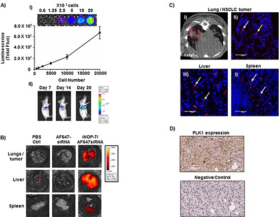
(A) Panel I, a representative image and graph showing increased luciferase activity with increasing H1299-Luc NSCLC cell numbers, n = 3 experiments. Panel II, pseudocolor images of mice injected orthotopically with H1299-Luc cells showing an increase in lung bioluminescence at days 7-20 post-tumor cell injection. (B) Representative ex-vivo fluorescent images of lungs and lung tumors, liver and spleen collected from mice with H1299-Luc orthotopic lung tumors, 4h post-injection (intravenous) with fluorescent siRNA (Red) complexed to iNOP-7. Mice injected with iNOP-7-fluorescent siRNA showed high fluorescence in the lung and lung tumors, liver and spleen. Mice injected with fluorescent siRNA alone or PBS served as controls. (C) Panel I, micro-CT image (axial) showing the presence of a growing tumor within the lungs of a mouse prior to injection with iNOP-7-fluorescent siRNA (red-dotted line marks the area of the tumor within the lung). Panels II-IV, are representative confocal microscopy images of frozen sections of lung tumor, liver and spleen showing the presence of fluorescent siRNA when delivered by iNOP-7, 4h post-injection (white arrows mark the location of fluorescent siRNA). (D) Panel I, Immunohistochemical image demonstrating PLK1 expression in orthotopic H1299-Luc tumors growing in the lung of mice. Panel II, immunohistochemical image of the corresponding antibody isotype control.