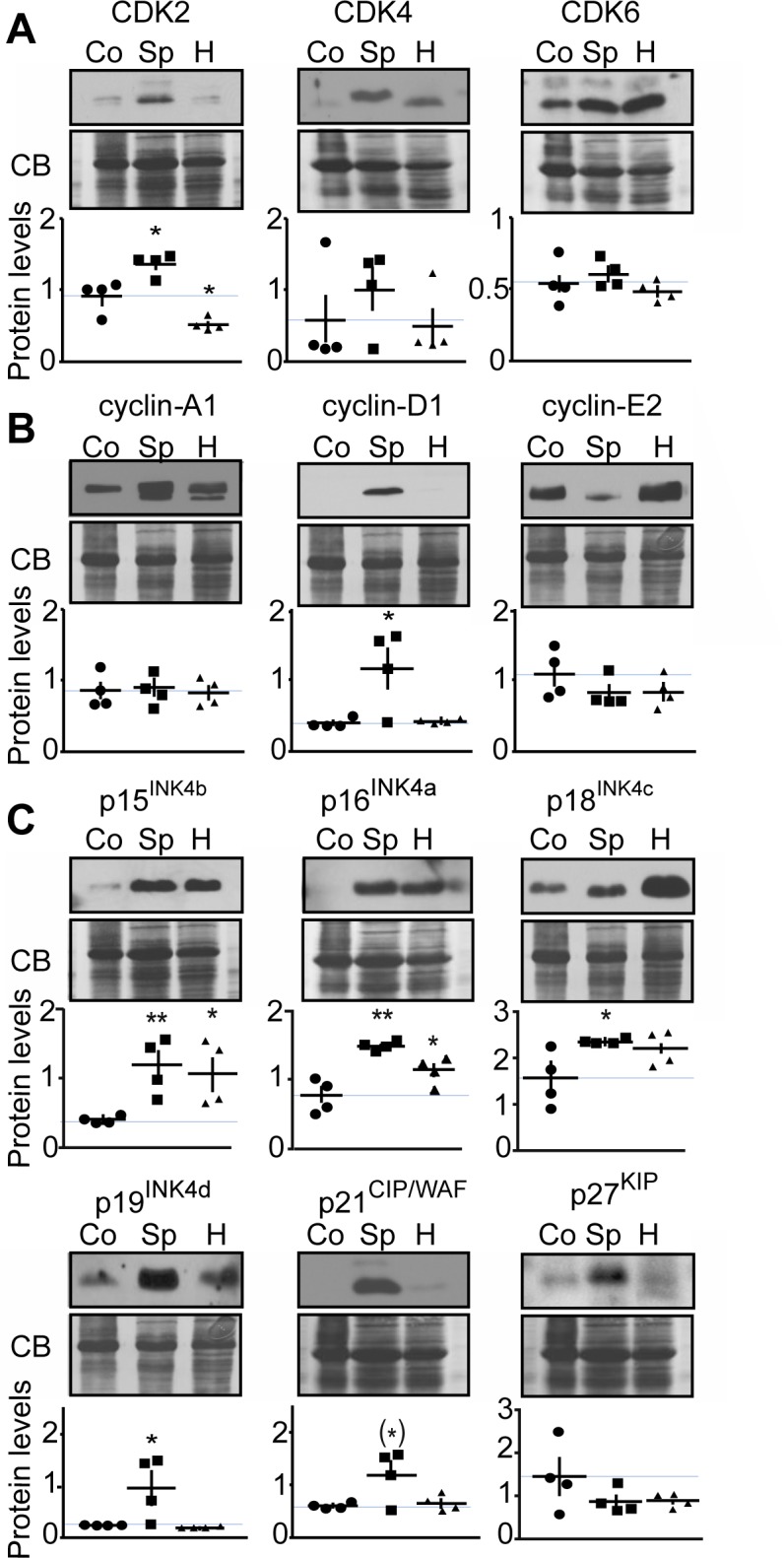
Figure 5. Analysis of cell cycle protein expression in human MTC samples.

Representative immunoblots of lysates from control thyroid tissue (Co), sporadic (Sp) and hereditary (H) human MTC tumors with antibodies as indicated are shown with quantification. Protein levels were normalized to Coomassie blue (CB) signal. Immunoblots were probed with antibodies to A) CDK2, CDK4 and CDK6; B) cyclins-A1, -D1, -E2; C) in p15INK4b, p16INK4a, p18INK4c, p19INK4d, p21CIP/WAF1 and p27KIP. P-values were for CDK2, Sp, p = 0.0168 and H, p = 0.0140; for CDK4, Sp, p = 0.4072 and H, p = 0.8576; for CDK6, Sp, p = 0.5484 and H, p = 0.4916; for cyclin-A1, Sp, p = 0.9923 and H, p = 0.7017; for cyclin-D1, Sp, p = 0.0307 and H, p = 0.6883; for cyclin-E2, Sp, p = 0.2645 and H, p = 0.3070; for p15INK4b, Sp, p = 0.0088 and H, p = 0.0310; for p16INK4a, Sp, p = 0.0014 and H, p = 0.0496; for p18INK4c, Sp, p = 0.0309 and H, p = 0.1054; for p19INK4d, Sp, p = 0.0484 and H, p = 0.3560; for p21CIP/WAF, Sp, p = 0.0554 and H, p = 0.6807; for p27KIP1, Sp, p = 0.2149 and H, p = 0.2131. Data are represented as mean +/− SEM, N = 4 for each condition.
