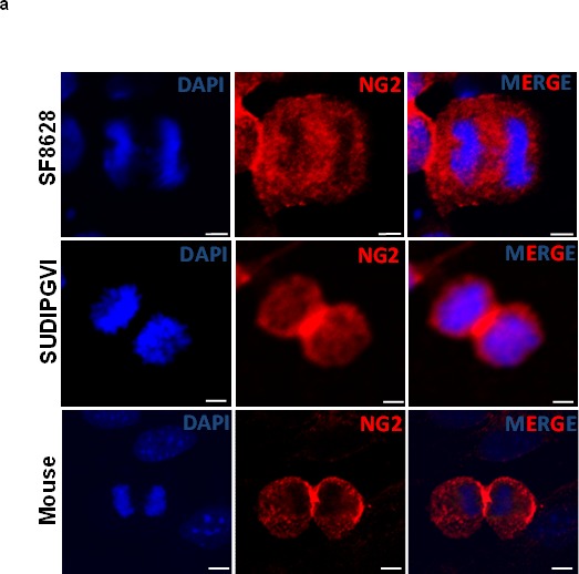
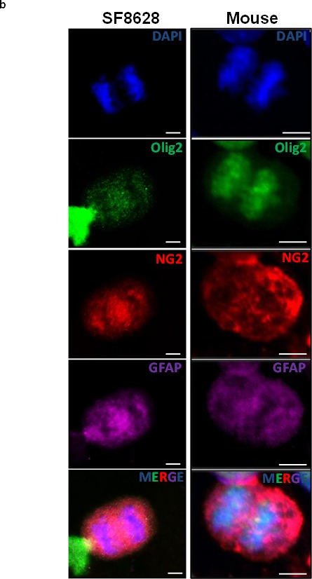
Figure 4. Mouse and human DIPG neurospheres exhibit symmetric NG2 expression in vitro and are immature stem cells co-expressing oligodendrocyte (Olig2) and astrocyte (GFAP) markers.


(a) PDGFB mouse tumor neurospheres (Mouse) and human primary DIPG cells (SF8628 and SUDIPGVI) were grown and stained using NG2 antibody (red) and DAPI (blue) nuclear staining. NG2 is equally expressed in dividing cells suggesting that its expression is defective (symmetric) in mitotic cells. Scale bar = 5 μm. (b) PDGFB mouse tumor (Mouse) and SF8628 human DIPG primary cells were expanded in culture and immunostained for DAPI (nuclear), oligodendrocyte marker (Olig2, green), NG2 (red), and astrocyte marker (GFAP, magenta). Confocal microscopy was used to assess expression of above-mentioned proteins in mitotic cells. Scale bar = 5 μM.
