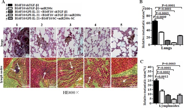
Figure 6. Histological analysis of lungs and lymph nodes of melanoma bearing mice.

A. Tissue sections derived from melanoma bearing mice 51 days after vaccination followed by challenge with the differently treated cells. The top and bottom panels show the lung sections and the lymph node sections, respectively. The arrows point to the metastatic focus as described in the text. B and C. Quantitative analysis of the tumor metastatic rates of lungs and lymph nodes, respectively, in the differently treated mice; refer to the statistical differences as indicated.
