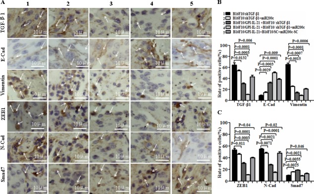
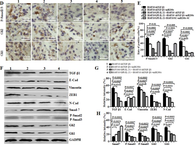
Figure 7. EMT-associated molecule expression analyzed by immunohistochemistry and Western blot in vaccinated mice challenged with B16F10/shTGF-β1 cells plus minus miR200c agomir.


A and D. Images shows the immunohistochemistry results. The arrows point to the EMT associated molecular expressions. B, C and E. Statistical analysis of the positive cells expressed with EMT associated molecules. F. EMT associated molecular expression analyzed by the Western blot assay. G and H. Semi-quantification analysis of molecular expression; refer to the statistical differences as indicated.
