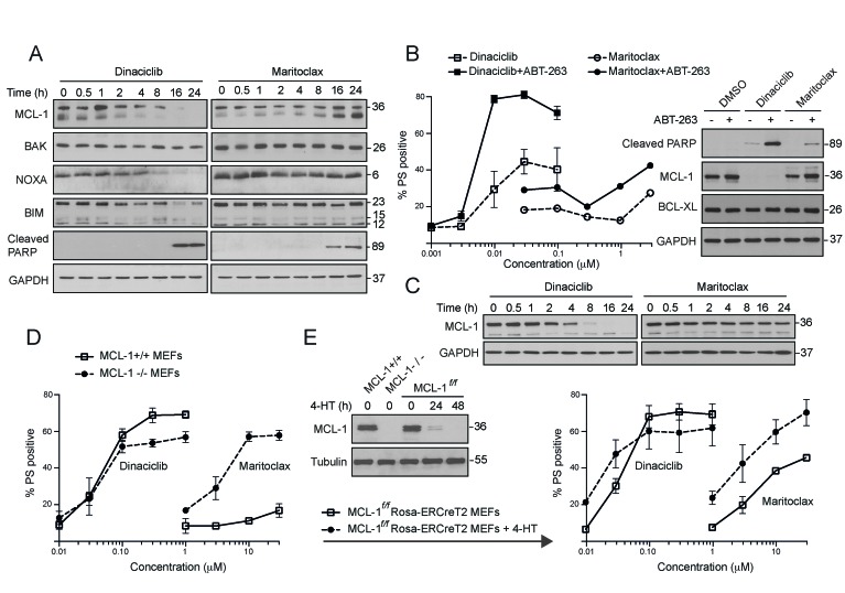
Figure 2. Dinaciclib and maritoclax diminish MCL-1 expression levels and induce MCL-1-dependent apoptosis in a cell-type specific manner.
(A) Whole cell lysates isolated from H460 cells, exposed to dinaciclib (30 nM) or maritoclax (3 μM) for the indicated times, were immunoblotted with the indicated antibodies. (B) H1299 cells were exposed to DMSO or ABT-263 (5 μM) for 30 min, followed for a further 24 h by the indicated concentrations of dinaciclib or maritoclax and cell death assessed by PS externalization. Western blots reveal changes in MCL-1 expression and PARP cleavage in H1299 cells, following 24 h of exposure to dinaciclib (30 nM) or maritoclax (3 μM), with or without a 30 min pretreatment of ABT-263 (5 μM). (C) Whole cell lysates from H1299 cells exposed to dinaciclib (30 nM) or maritoclax (3 μM) for the indicated times were immunoblotted with the indicated antibodies. (D) MEFs deficient in MCL-1 (dashed lines) along with their wild type counterparts (continuous bold lines) were exposed for 24 h to the indicated inhibitors and apoptosis assessed by PS externalization. (E) MCL-1f/fRosa-ERCreT2 MEFs were initially exposed to DMSO or 4-hydroxytamoxifen (4-HT) (100 nM) to delete endogenous MCL-1. The cells were then exposed for a further 24 h to the indicated inhibitors and apoptosis assessed. Western blots reveal changes in MCL-1 expression in different MEFs, following 4-HT exposure for 0, 24 or 48 h. Error bars represent the Mean ± SEM from three independent experiments. In all the graphs, the extent of apoptosis in untreated control cells matched the % apoptosis of the lowest concentration tested for both inhibitors.

