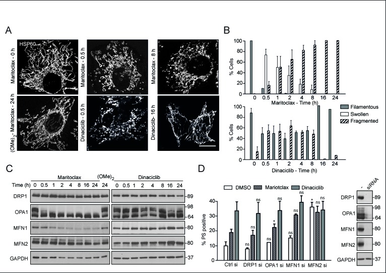
Figure 4. Maritoclax, and dinaciclib induce marked mitochondrial structural changes, which may contribute to apoptosis.
(A) H460 cells, grown on coverslips, were exposed for different times to dinaciclib (30 nM), maritoclax (3 μM) or dimethoxymaritoclax (3 μM), stained with antibody against HSP60 and subjected to confocal microscopy. Scale bar – 10 μm. (B) At least 200 cells from five different fields were quantified for changes in mitochondrial morphology (categorised as filamentous, swollen or fragmented) and a graph plotted from three independent replicates. Error bars represent the Mean ± SEM. (C) Whole cell lysates from H460 cells, exposed to of dinaciclib (30 nM), maritoclax (3 μM) or dimethoxymaritoclax (3 μM) for the indicated times, were immunoblotted with the indicated antibodies. (D) H460 cells, reverse-transfected with the indicated siRNA oligoduplexes for 48 h, were exposed for a further 24 h to dinaciclib (30 nM) or maritoclax (3 μM), and cell death assessed by PS externalization. Error bars represent the Mean ± SEM from three independent experiments. Western blots show the knockdown efficiency of the individual siRNAs. Statistical analysis was conducted using a paired t-test (*P < 0.05, ns – not significant, if P > 0.05).

