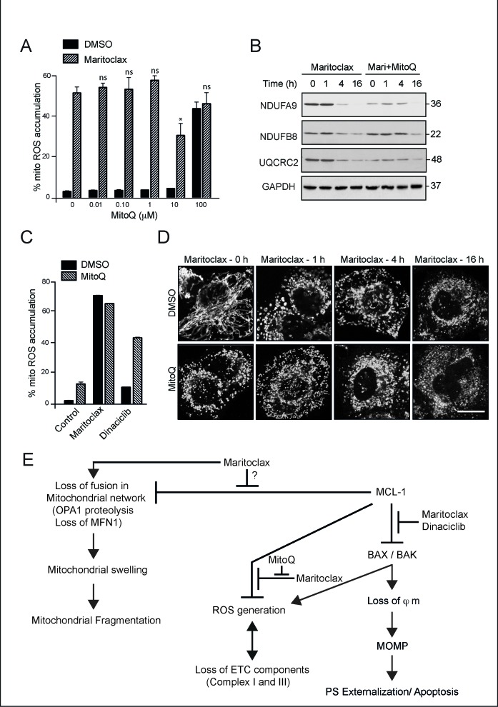
Figure 7. Maritoclax-mediated mitochondrial effects are partially rescued by MitoQ.
(A) H460 cells were exposed to different concentrations of MitoQ for 4 h, followed by maritoclax (3 μM) for a further 2 h, and mitochondrial accumulation of ROS assessed. Statistical analysis was conducted using a paired t-test (*P < 0.05, ns – not significant, if P > 0.05). (B) Whole cell lysates from H460 cells, exposed to MitoQ (10 μM) for 4 h followed by maritoclax (3 μM) for the indicated times, were immunoblotted with the indicated antibodies. (C) H460 cells exposed to MitoQ (10 μM) for 4 h, followed by either Maritoclax (3 μM) or dinaciclib (30 nM) for a further 20 h were stained with MitosoxRed to monitor changes in mitochondrial ROS accumulation. Error bars represent the Mean ± SEM from three independent experiments. (D) H460 cells, grown on coverslips, were exposed for 4 h to MitoQ (10 μM), followed by maritoclax (3 μM) for the indicated times. The cells were then immunostained with HSP60 antibody and subjected to confocal microscopy. Scale bar – 10 μm. (E) Scheme for proposed mechanism of action of dinaciclib and maritoclax. Dinaciclib and maritoclax interfere with the anti-apoptotic effects of MCL-1, in a Bax/Bak and caspase-9-dependent manner, by causing the loss of mitochondrial membrane potential (φm) and activation of mitochondrial outer membrane permeabilization (MOMP), resulting in apoptosis. Maritoclax, but not dinaciclib, interfered with other functions of MCL-1, including the postulated regulation of mitochondrial fusion, accompanied by enhanced OPA1 proteolysis and loss in MFN1, all of which resulted in mitochondrial structural changes that initially manifested as swelling and subsequently resulted in extensive mitochondrial fragmentation. However, maritoclax-mediated changes in mitochondrial structure also occurred in cells lacking MCL-1, suggesting the involvement of MCL-1-independent mechanisms in these processes. Maritoclax also resulted in ROS generation in mitochondria, which accompanied a loss in several components of the electron transport chain, in a Bax/Bak-dependent but a MCL-1-independent manner, thus placing Bax/Bak upstream to mitochondrial ROS induction and the loss of ETC components.

