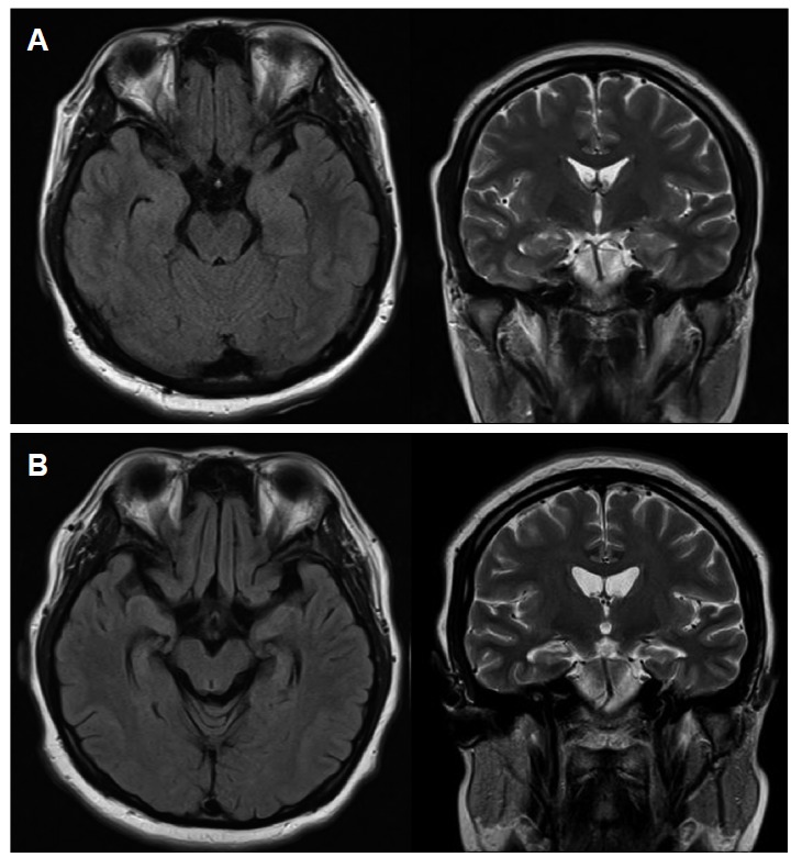
Figure 1.

Brain MRI. (A) Initial brain MRI was normal. (B) Follow-up MRI, ten months after the onset of symptoms, shows diffuse brain atrophy and bilateral hippocampal atrophy compared with the initial brain MRI (A) on fluid attenuation inversion recovery (FLAIR) and T2 weighted images (T2WI).
