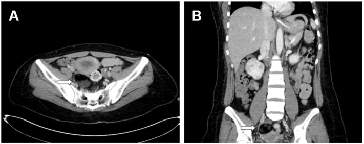
Figure 2.

Abdomen-Pelvic CT. (A) The axial abdomen-pelvic CT shows a large cystic mass (4.8 × 5.0 cm, white arrow) in the right adnexa. (B) The coronal abdomen-pelvic CT shows a large cystic mass (white arrow), which was confirmed to be a mature cystic teratoma.
