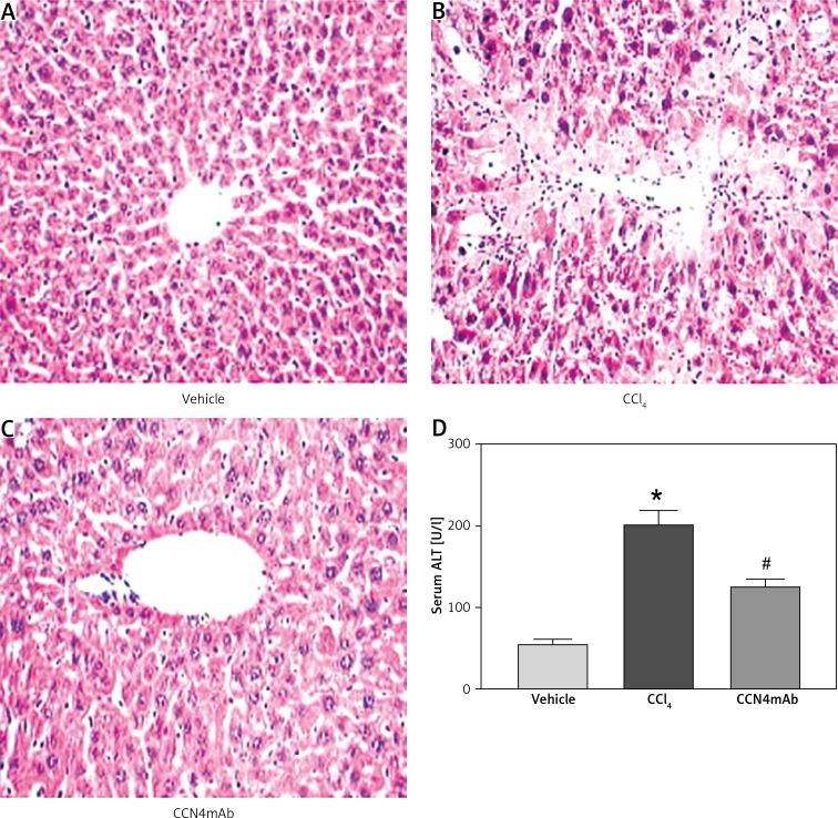
Figure 2.
Representative photographs of HE staining: A – vehicle-treated normal control, B – CCl4-treated and C – CCl4 + CCN4mAb-treated groups. Liver necrotic area was present in CCl4-treated mice with significantly large amounts of inflammatory cell infiltration surrounding the centrilobular veins. CCl4 treatment resulted in obviously increased (D) serum ALT level
*p < 0.01, vs. vehicle-treated normal control; #p < 0.01 vs. CCl4-treated group (n = 10–12/group). Magnification: 200×.

