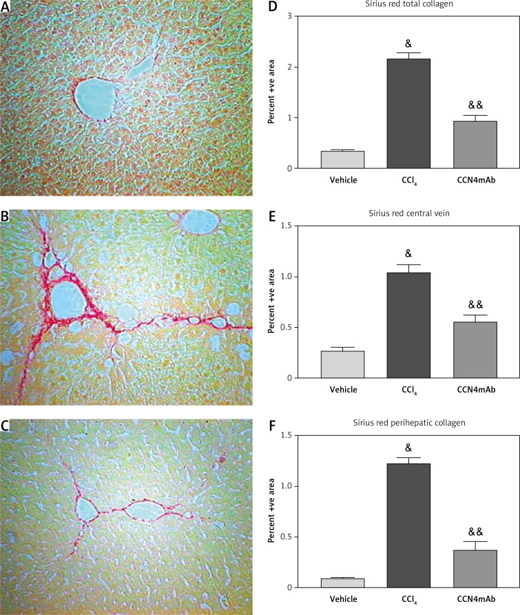
Figure 3.
Sirius Red staining on the sections of (A) vehicle-treated normal control, (B) CCl4-treated and (C) CCl4 + CCN4mAb-treated groups. Histograms show the percentage area of Sirius Red staining of collagen of (D) whole liver section, (E) along central vein and (F) pericellular area. CCl4 treatment increased collagen deposition in central vein and pericellular area
&p < 0.01, versus vehicle-treated normal control; &&p < 0.01 compared with CCl4-treated group (n = 10–12/group).

