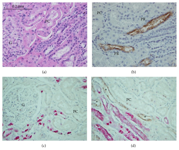
Figure 1.
Acute tacrolimus nephrotoxicity, identified by KIM-1 staining in the case. On routine light microscopy, the renal tissue appears normal in pars convoluta (PC) and medullary rays located in the right lower corner of A (H&E staining). However, KIM-1 staining (B) revealed 2+ positive staining (brown color) reflective of acute kidney injury in the proximal tubules located in medullary rays (pars recta, PR) but not in the proximal tubules around glomeruli (PC), indicating acute tubular injury, a pattern consistent with acute nephrotoxicity of tacrolimus. KIM-1 and cytokeratin-7 (a distal tubular marker) coexpression is shown in C and D. In both panel C and panel D, pink stained tubules were distal nephron tubules. In panel C, PC around glomerulus stained negatively for KIM-1 and in panel D, PR in medullary rays stained positively for KIM-1 (brown color staining, indicated by asterisk). Magnification ×400 for A-B and ×200 for C-D.

