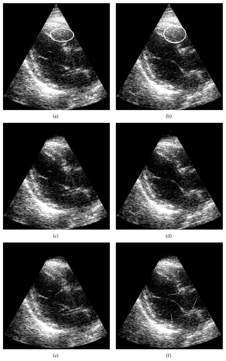
Figure 12.
Two examples of an apical view of heart images are illustrated before filtering (a, b) and after clutter removal with MCA (c, d) and after applying SVF (e, f). Ellipses in the unfiltered images indicate regions of clutter artifacts due to multipath reverberations. The arrows point to regions of tissue incorrectly filtered. Images are shown on a log compressed linear gray scale mapping to 0 to 30 dB.

