FIG 4.
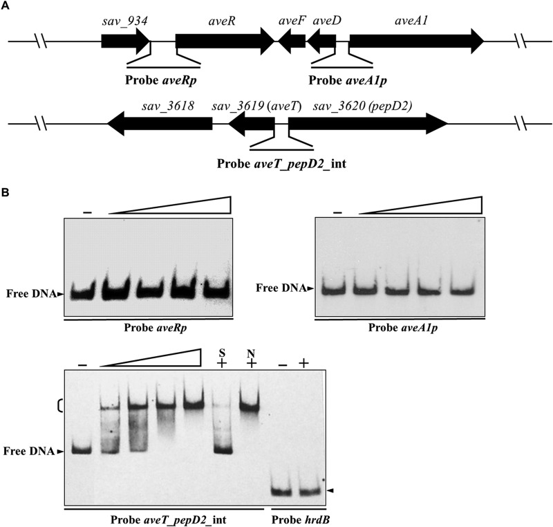
EMSAs of AveT binding to the aveT-pepD2 intergenic region. (A) Schematic representation of the probes used for EMSAs. Probe aveRp, 527-bp DNA fragment from positions +49 to −478 relative to the start codon of aveR; probe aveA1p, 333-bp DNA fragment from positions −6 to −338 relative to the start codon of aveA1; probe aveT_pepD2_int, a 248-bp DNA fragment covering the aveT-pepD2 intergenic region. Probes aveRp and aveA1p cover the putative TSSs of aveR and aveA1, respectively. (B) EMSAs of the interaction of probes aveRp, aveA1p, and aveT_pepD2_int with purified His6-AveT protein. Each reaction mixture contained 0.15 nM labeled probe. EMSAs with 300-fold unlabeled specific probe (lane S) or nonspecific competitor DNA (lane N) were performed to confirm the specificity of the band shifts. Labeled probe hrdB was used as a negative control. Labeled probes were incubated in the absence (lanes −) or presence of various amounts of His6-AveT. The concentrations of the His6-AveT protein for the probes were as follows: for aveRp and aveA1p, 0.4, 1.2, 2.0, and 2.8 μM; for aveT_pepD2_int, 0.005, 0.01, 0.05, and 0.1 μM; for hrdB, 2.8 μM. Competition experiments were performed using 0.1 μM His6-AveT. Arrowheads, free probe; bracket, AveT-DNA complex.

