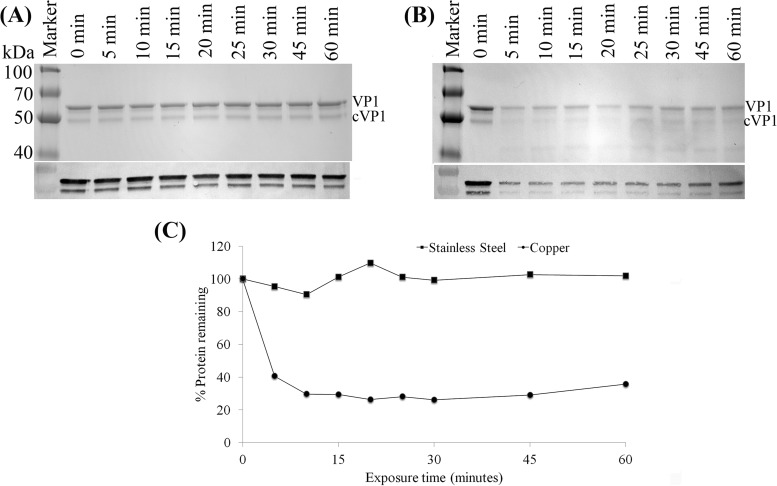
FIG 3.
Analysis of HuNoV capsid protein by SDS-PAGE and Western blotting. (A and B) One-microliter aliquots (representing 590 ng) of purified HuNoV GII.4 Houston VLPs were placed onto either stainless steel (A) or copper (B) surfaces and eluted at various time points in PBS-EDTA. Eluted VLPs were analyzed by 12% SDS-PAGE followed by protein staining. VP1, native full-length capsid protein; cVP1, cleaved VP1. Blots from Western blot analysis of VP1 proteins using mouse monoclonal antinorovirus GII.4 antibody are shown below each SDS-PAGE gel image. (C) Quantitative analysis of the remaining VP1 proteins detected by SDS-PAGE after surface exposure.

