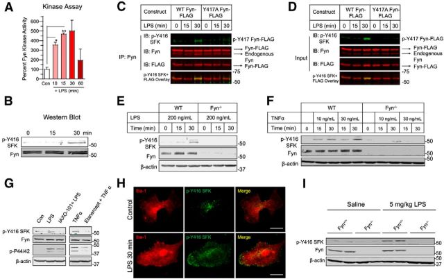
Figure 2.
Fyn kinase is rapidly activated in microglial cells and in the ventral midbrain following inflammogen stimulation. A, Fyn kinase assay shows that Fyn activity was highly induced in BV2 microglia treated with 1 μg/ml LPS for 10, 15, and 30 min. *p < 0.05. **p < 0.01. B, Immunoblots showing a concomitant rise in p-Y416 SFK levels in BV2 cell lysates after LPS treatment. C, D, Immunoprecipitation studies revealed that WT Fyn, but not activation loop tyrosine-mutant Fyn (Y417A Fyn), when overexpressed in BV2 microglia, was activated following LPS stimulation. E, F, Treatment of (E) primary microglia with LPS and TNFα (F) for 15 and 30 min increased p-Y416 SFK levels in primary microglia obtained from Fyn+/+, but not Fyn−/− mice, identifying Fyn as the primary Src family kinase that was activated by inflammogen stimulation. G, Pretreatment of primary microglia with the TLR-signaling antagonist IAXO-101 or the TNFα receptor decoy etanercept abolished Fyn activation by LPS or TNFα stimulation (p44/42 phosphorylation used as marker for early microglial activation). H, Immunocytochemistry of LPS-treated WT primary microglia showing that activated Fyn expression greatly increased and was localized preferentially to the membrane periphery of the microglial cell. Scale bar, 20 μm. I, Immunoblots of ventral midbrain lysates showed that peripheral administration of the inflammogen LPS (5 mg/kg) increased p-Y416 SFK levels in Fyn+/+, but not in Fyn−/− ventral midbrain tissues.

